

Leica DMi8 Inverted Microscope
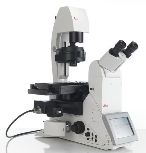
The Leica DMi8 is a modular, inverted research microscope platform engineered for flexibility in life science imaging. Built on an open-architecture stand, it allows for the integration of manual, coded, or fully motorized components depending on experimental requirements.
For advanced applications, the system supports THUNDER technology, which utilizes Computational Clearing to remove out-of-focus blur from widefield images in real time, enabling high-contrast 3D imaging of thick tissues. Stability for long-term time-lapse experiments is maintained via Adaptive Focus Control (AFC), a hardware-based LED feedback loop that actively corrects thermal Z-drift.
Technical Specification of Leica DMi8
Technical Specifications
Optional: AFC (Adaptive Focus Control) Hardware Drift Correction
Key Features and Benefits of Leica DMi8
Uses proprietary Computational Clearing to eliminate out-of-focus blur in real time, delivering sharp, high-contrast images of thick 3D samples without the need for a confocal system.
- Resolve fine details in thick tissue sections instantly.
- Works with standard GFP/mCherry fluorophores.
A unique physical access point to the microscope's infinite light path. It allows you to couple additional light sources, lasers (for FRAP/photoactivation), or custom optical devices directly into the system.
- Future-proof your lab for new techniques.
- Mix multiple illumination methods seamlessly.
Automatically dispenses the correct amount of water immersion medium during experiments. It compensates for evaporation or stage movement, enabling long-term high-NA imaging.
- Perform 48+ hour time-lapses with water objectives.
- Maintains physiological conditions for cells.
Simply place your sample on the stage, and the system automatically scans the holder to generate a low-mag overview map. Click anywhere on the map to move directly to that region.
- Eliminates tedious manual searching.
- Identify transfected cells in seconds.
An LED-based hardware autofocus system that actively monitors the distance to the coverslip in real time, correcting for thermal drift immediately.
- Keeps cells perfectly in focus during long incubations.
- Works with all contrast methods and objectives.
Applications and Usecases of Leica DMi8 Inverted Microscope
Monitor cell division, migration, and differentiation over days without losing focus.
- AFC prevents thermal drift during temperature cycles.
- Adaptive Immersion enables high-NA water objectives for days.
Automate the imaging of multi-well plates (96/384) to generate statistically significant data sets.
- Large 19/22mm FOV captures more cells per image.
- Sample Finder maps the entire plate instantly.
Stable, vibration-free platform for precise injection (ICSI) or patch-clamp electrophysiology.
- Open stage design allows easy access for manipulators.
- Integrated Modulation Contrast (IMC) for clear viewing.
Expand the system with GSD or TIRF modules for single-molecule localization.
- Infinity Port enables easy laser coupling.
- Nanometer-precision stage for localization microscopy.
Frequently Asked Questions
Yes. The DMi8 is built on an **open platform** concept. You can start with a manual configuration and upgrade later to include motorized stages, fluorescence turrets, AFC focus control, or even super-resolution modules like TIRF or GSD.
The **Infinity Port** is a specialized access point into the microscope's fluorescence light path. It allows researchers to couple additional optical devices—such as pulsed lasers for FRAP, photoactivation units, or custom light sources—without interfering with the standard camera or eyepiece paths.
Absolutely. The **Scanning Stage** option is designed for high-speed movement across 96, 384, or even 1536-well plates. Combined with the **Automated Sample Finder**, you can map and image entire plates in minutes.
AFC uses an infrared LED beam that reflects off the interface between the coverslip and the media. A sensor detects any shift in this reflection (caused by thermal expansion or drift) and instantly adjusts the Z-drive to maintain perfect focus.
Confocal microscopes use pinholes to physically block out-of-focus light (slow, high phototoxicity). **THUNDER** uses widefield illumination (fast, gentle) combined with **Computational Clearing** to mathematically remove the blur. This gives confocal-like images at much higher speeds.
Yes, by using the **Adaptive Immersion** module. This system automatically dispenses water immersion medium to the objective tip during the experiment, compensating for evaporation so you don't lose image quality over time.
The DMi8 is fully integrated with **Leica LAS X (Leica Application Suite X)**. This software handles everything from basic image capture to complex multidimensional acquisition, THUNDER processing, and analysis.
The standard camera port offers a **19 mm FOV**. However, the system optics can support up to a **22 mm FOV** when paired with specific sCMOS cameras, allowing you to capture significantly more data per image tile.